
Axial skeleton
1 Frontal bone
2 Occipital bone
3 Parietal bone
4 Orbit
5 Nasal cavity
6 Maxilla
7 Zygomatic bone
8 Mandible

Appendicular sketelton
Upper limb and shoulder girdle 19 Clavicle
20 Scapula
21 Humerus
22 Radius
23 Ulna
24 Carpal bones
25 Metacarpal bones
26 Phalanges of the hand
Lower limb and pelvis
27 Ilium
28 Pubis
29 Ischium
30 Symphysis pubis
31 Femur
32 Tibia
33 Fibula
34 Patella
35 Tarsal bones
36 Metatarsal bones
37 Phalanges of the foot
38 Calcaneus

1 Coronal suture
2 Frontal bone
3 Sphenoidal bone
4 Sphenofrontal suture
5 Ethmoidal bone
6 Nasal bone
7 Nasomaxillary suture
8 Lacrimal bone
9 Lacrimomaxillary suture
10 Ethmoidolacrimal suture
11 Zygomatic bone
12 Anterior nasal spine
13 Maxilla
14 Mandible
15 Mental foramen
16 Mental protuberance
17 Superior temporal line
18 Inferior temporal line
19 Parietal bone
20 Temporal bone
21 Squamous suture
22 Lambdoid suture
23 Temporal fossa
24 Parietomastoid suture
25 Occipital bone
26 Zygomatic arch
27 Occipitomastoid suture
28 External acoustic meatus
29 Mastoid process
30 Tympanic portion of temporal bone
31 Condylar process of mandible
32 Coronoid process of mandible

1. Sinus Frontalis
2 Cellulae ethmoidales
3 Sinus Sphenoidalis
4 Concha nasalis superior
5 Concha nasalis media
6 Hiatus Maxillaris
7 Concha nasalis inferior
8 Os Palatina
9 Maxilla
10 Meatus inferior
11 Processus palatinus

OS FRONTALIS
1. Squama frontalis
2. Linea temporalis inferior
3. facies temporalis (ossis frontalis)
4. Foramen supraorbitale
5. Processus zygomaticus
OS OCCIPITALIS
6. Squama occipitalis
OS SPHENOIDALE
7. ala major ossis sphenoidalis
8. canalis opticus / foramen opticum ossis sphenoidalis
9. lamina lateralis processus pterygoidei
OS PALATINUM
10. processus orbitalis
11. lamina perpendicularis
12.Crista conchalis
13. Crista nasalis
14. Lamina horizontalis
OS ETHMOIDALE
15. Lamina orbitalis
16. Cellulae ethmoidales
17. Concha medialisa
18. Lamina Perpendicularis
OS MAXILLA
19. Sulcus infraorbitalis
20. Foramen infraorbitalis
21. Processus zygomaticus
22. processus alveolaris
23. Processus palatina
CONCHAE NASALIS INFERIOR SINISTRA
24. Concha inferior

1 Fissura orbitalis superior
2 Foramen rotundum
3 Canalis opticus
6 Meatus acusticus internus
7 Foramen Jugularis
15 Crista galli
16 Lamina cribrosa
17 Impressiones digitatae (Os Frontalis)
18 Ala minor ossis sphenoidalis
19 Foramen lacerum
20 Hypophyseal fossa
(sella turcica)
21 Processus clinoideus anterior
22 Impressio trigeminalis
23 Pars petrosa ossis temporalis
24 sulcus sinus sigmoidei ossis parietalis
25 Dorsum sellae
(posterior clinoid process)
26 ala major ossis sphenoidalis, Sulcus arteriae meningeae mediae
27 canalis hypoglossi

2 Foramen cecum
3 Crista galli
4 Cribriform plate of ethmoidal bone
5 Lesser wing of sphenoidal bone
6 Superior orbital fissure
7 Foramen rotundum
8 Carotid sulcus
9 Middle cranial fossa
10 Foramen ovale
11 Foramen spinosum
12 Clivus
13 Groove for superior petrosal sinus
14 Jugular foramen
15 Groove for sigmoid sinus
16 Internal occipital crest
17 Groove for transverse sinus
18 Internal occipital protuberance
19 Digitate impressions
20 Anterior cranial fossa
21 Chiasmatic sulcus
22 Anterior clinoid process
23 Optic canal
24 Sella turcica (hypophysial fossa)
25 Posterior clinoid process
26 Dorsum sellae
27 Foramen lacerum
28 Groove for greater petrosal nerve
29 Internal acoustic meatus
30 Hypoglossal canal
31 Foramen magnum
32 Posterior cranial fossa
33 Diploe

3 Sutura sagitalis
6 Parietal foramen
7 Parietal tuber or eminence
14 Lambdoid suture
15 Os Occipitalis
16 External occipital protuberance
17 Inferior nuchal line
18 sutura occipitomastoid
19 Os Temporalis
20 Processus Mastoideus
21 Incisura mastoidea

1 Incisive canal
2 Median palatine suture
3 Palatine process of maxilla
4 Palatomaxillary suture
5 Greater and lesser palatine foramina
6 Inferior orbital fissure
7 Middle concha (process of ethmoidal bone)
8 Vomer
9 Foramen ovale
10 Groove for auditory tube
11 Pterygoid canal
12 Styloid process
13 Carotid canal
14 Stylomastoid foramen
15 Jugular foramen
16 Groove for occipital artery
17 Occipital condyle
18 Condylar canal
19 Nuchal plane
20 External occipital protuberance
21 Zygomatic arch
22 Lateral pterygoid plate
23 Medial pterygoid plate
24 Mandibular fossa
25 Pharyngeal tubercle
26 Superior nuchal line
27 Mastoid process
28 Inferior nuchal line
29 Mastoid notch
30 Foramen magnum

8 Supra-orbital margin
14 Squamous part of frontal bone
18 Zygomatic process of frontal bone
20 Maxilla
21 Frontal process of maxilla
22 Lacrimal bone (yellow)
23 Zygomatic bone (orange)
24 Zygomaticofacial foramen
Temporal bone
25 Squamous part of temporal bone
26 External acoustic meatus
27 Mastoid process
28 Styloid process
29 Mandibular fossa
30 Articular tubercle
31 Zygomatic process
Occipital bone
32 Squamous part of occipital bone

Os Clavicula
1 Acromial end
2 Articular facet for the acromion
3 Articular facet for the sternum
4 Sternal end
5 Trapezoid line
6 Conoid tubercle
7 Impression of costoclavicular ligament

Os Scapula
C = Lateral border
D = Superior angle
E = Inferior angle
1 Acromion
2 Coracoid process
4 Glenoid cavity
5 Infraglenoid tubercle
7 Spine
11 Supraglenoid tubercle
13 Base of coracoid process

Humerus
1 Greater tubercle
2 Lesser tubercle
3 Crest of lesser tubercle
4 Crest of greater tubercle
5 Intertubercular sulcus
6 Surgical neck
7 Deltoid tuberosity
8 Antero-lateral surface
9 Lateral supracondylar ridge
10 Radial fossa
11 Lateral epicondyle
12 Capitulum
13 Head
14 Anatomical neck
15 Antero-medial surface
16 Medial supracondylar ridge
17 Coronoid fossa
18 Medial epicondyle
19 Trochlea
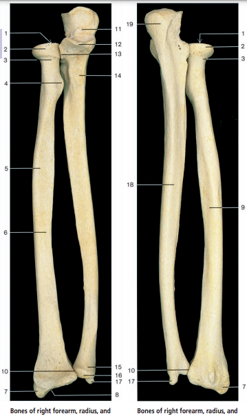
Radius
1 Head
2 Articular circumference
3 Neck
4 Radial tuberosity
5 Shaft
6 Anterior surface
7 Styloid process
8 Articular surface
9 Posterior surface
10 Ulnar notch
Ulna
11 Trochlear notch
12 Coronoid process
13 Radial notch
14 Ulnar tuberosity
15 Head
16 Articular circumference
17 Styloid process
18 Posterior surface
19 Olecranon
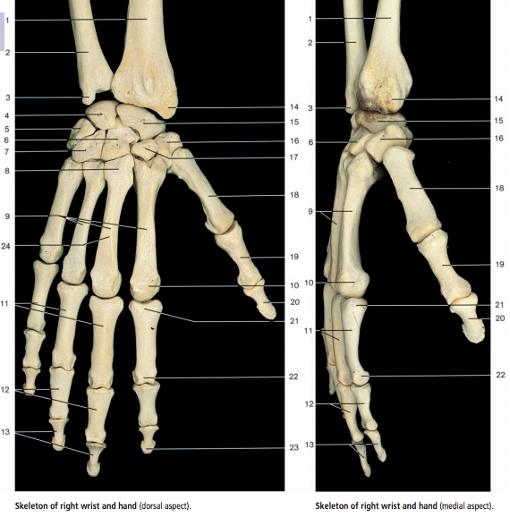
1 Radius
2 Ulna
3 Styloid process of ulna
4 Lunate bone
5 Triquetral bone
6 Capitate bone
7 Hamate bone
8 Base of third metacarpal bone
9 Metacarpal bones
10 Head of metacarpal bone
11 Proximal phalanges of the hand
12 Middle phalanges of the hand
13 Distal phalanges of the hand
14 Styloid process of radius
15 Scaphoid bone
16 Trapezium bone
17 Trapezoid bone
18 Metacarpal bone of thumb
19 Proximal phalanx of thumb
20 Distal phalanx of thumb
21 Base of second proximal phalanx
22 Head of second
proximal phalanx
23 Tuberosity
of distal phalanx
24 Body of third
metacarpal bone

1 Radius
2 Styloid process of radius
3 Scaphoid bone
4 Capitate bone Carpal bones 5 Trapezium
6 Trapezoid bone
7 First metacarpal bone
8 Second to fourth metacarpal bones
9 Proximal phalanx of thumb
10 Distal phalanx of thumb
11 Base of second proximal phalanx
12 Proximal phalanges
13 Head of second proximal phalanx
14 Middle phalanges
15 Distal phalanx
16 Ulna
17 Styloid process of ulna
18 Lunate bone
19 Pisiform bone
20 Triquetral bone Carpal bones
21 Hamate bone
22 Hamulus or hook
of hamate bone
23 Base of third metacarpal bone
24 Head of metacarpal bone
25 Tuberosity of distal phalanx


1 Lateral condyle of tibia
2 Position of tibiofibular joint
3 Head of fibula
4 Interosseous border of tibia
5 Shaft of fibula
6 Interosseous border of fibula
7 Lateral surface of fibula
8 Position of tibiofibular syndesmosis
9 Lateral malleolus
10 Medial condyle of tibia
11 Tuberosity of tibia
12 Shaft of tibia (diaphysis)
13 Anterior margin of tibia
14 Medial malleolus
15 Inferior articular surface
of tibia
16 Intercondylar eminence
17 Soleal line
18 Medial border of tibia
19 Posterior surface of tibia
20 Malleolar sulcus of tibia
21 Malleolar articular surface
of fibula
22 Apex of head of fibula
23 Posterior surface of fibula
24 Posterior border of fibula
25 Medial intercondylar tubercle
26 Posterior intercondylar area
27 Anterior intercondylar area
28 Lateral intercondylar tubercle

1 Tuberosity of distal phalanx of great toe
2 Distal phalanx of great toe
3 Proximal phalanx of great toe
4 Head of first metatarsal bone
5 First metatarsal bone
6 Base of first metatarsal bone
7 Medial cuneiform bone
8 Intermediate cuneiform bone
9 Position of cuneonavicular joint
10 Navicular bone
11 Position of talocalcaneonavicular joint
12 Head of talus
13 Neck of talus
14 Trochlea of talus
15 Posterior talar process
16 Distal phalanges
17 Middle phalanx
18 Position of interphalangeal joints
19 Proximal phalanges
20 Position of metatarsophalangeal joints
21 Metatarsal bones
22 Position of tarsometatarsal joints
23 Lateral cuneiform bone
24 Tuberosity of fifth metatarsal bone
25 Cuboid bone
26 Position of calcaneocuboid joint
27 Calcaneus
28 Tarsal sinus
29 Lateral malleolar surface of talus
30 Peroneal trochlea of calcaneus
31 Groove for the tendon of peroneus longus muscle
32 Calcaneal tuberosity
33 Sustentaculum tali
34 Tibia
35 Medial malleolus
36 Fibula
37 Position of tibiofibular syndesmosis
38 Position of ankle joint
39 Lateral malleolus
40 Position of subtalar joint

A = Ilium
B = Ischium
C = Pubis
1 Posterior superior iliac spine
2 Posterior gluteal line
3 Posterior inferior iliac spine
4 Greater sciatic notch
5 Ischial spine
6 Lesser sciatic notch
7 Body of ischium
8 Ischial tuberosity
9 Obturator foramen
10 Iliac crest
11 Anterior gluteal line
12 Internal lip of iliac crest
13 External lip of iliac crest
14 Anterior superior iliac spine
15 Inferior gluteal line
16 Anterior inferior iliac spine
17 Lunate surface of acetabulum
18 Acetabular fossa
19 Acetabular notch
20 Pecten pubis
21 Pubic tubercle
22 Body of pubis
23 Iliac fossa
24 Arcuate line
25 Iliopubic eminence
26 Symphysial surface of pubis
27 Auricular surface
28 Pelvic surface of sacrum
29 Superior articular process of sacrum
30 Dorsal sacral foramina
31 Sacral tuberosity
32 Lateral sacral crest
33 Median sacral crest
34 Obturator groove
35 Coccyx
Comments
Post a Comment